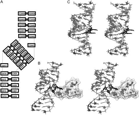
FIGURE 5.
(A) Secondary structure and (B) stereo view of ribosomal kissing-loop complex from H. marismortui 50S subunit with bulged-out bases (highlighted in black) forming tertiary contacts to the adjacent part of the 23S rRNA (unfilled surface). (C) Averaged MD structure over the last 10 ns with stacked bulges (black).

