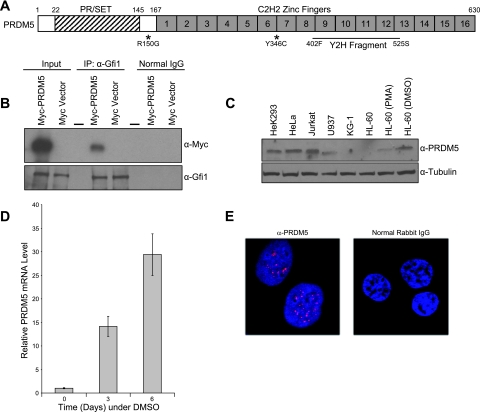
FIG. 1.
PRDM5 structure, Gfi1 interaction, neutropenic sequence variants, and subcellular localization. (A) PRDM5 schematic representation. Asterisks denote variants found in neutropenic individuals. Zinc finger fragment recovered in yeast two-hybrid (Y2H) screen with zinc fingers of Gfi1 is indicated. Numbers on top of schematic correspond to protein sequence. (B) Interaction of PRDM5 with Gfi1 in cotransfected HeLa cells. Coimmunoprecipitation assays were performed using an antibody against Gfi1 or normal goat IgG as a negative control with HeLa cells cotransfected with the indicated plasmids. Dash indicates empty lane. (C) Western blot analysis of PRDM5 in human cell lines. PMA, phorbol myristate acetate; DMSO, dimethyl sulfoxide. (D) Up-regulation of PRDM5 during dimethyl sulfoxide (DMSO)-induced granulocytic differentiation of HL-60 cells, as measured by quantitative real-time RT-PCR with GAPDH as internal control. (E) Confocal microscopy of indirect immunofluorescent staining of HeLa cells with PRDM5 antibody (red) and control IgG. Nuclei counterstained with DAPI are blue. α, anti.

