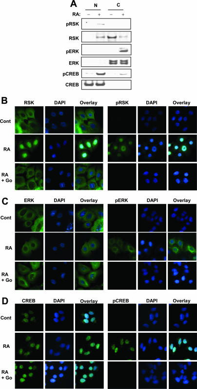
FIG. 7.
ERK and RSK are activated by RA, and RSK but not ERK is translocated to the nucleus. (A) NHTBE cells were preincubated with 10 μM Go6976 (selective PKCα inhibitor) for 2 h and then further treated with or without 1 μM RA for 30 min. Nuclear (N) and cytosolic (C) extracts were prepared as described in Materials and Methods. Equal amounts of proteins from each fraction were examined using Western blot analysis with the indicated antibodies. (B to D) Immunocytofluorescence analysis of the activation and translocation of RSK, ERK, and CREB. NHTBE cells grown in an RA-deficient medium on coverslips for 7 days were subjected to immunocytofluorescence analysis. Cells were preincubated with or without 10 μM Go6976 (Go) and then further incubated with a vehicle (Cont) (top) or 1 μM RA (middle) for 30 min before fixation. After fixation, the cells were stained with the indicated antibodies followed by anti-mouse AlexaFluor 488 antibodies (green). Nuclei were stained with DAPI (blue) and then merged. All figures are representative of three independent experiments.

