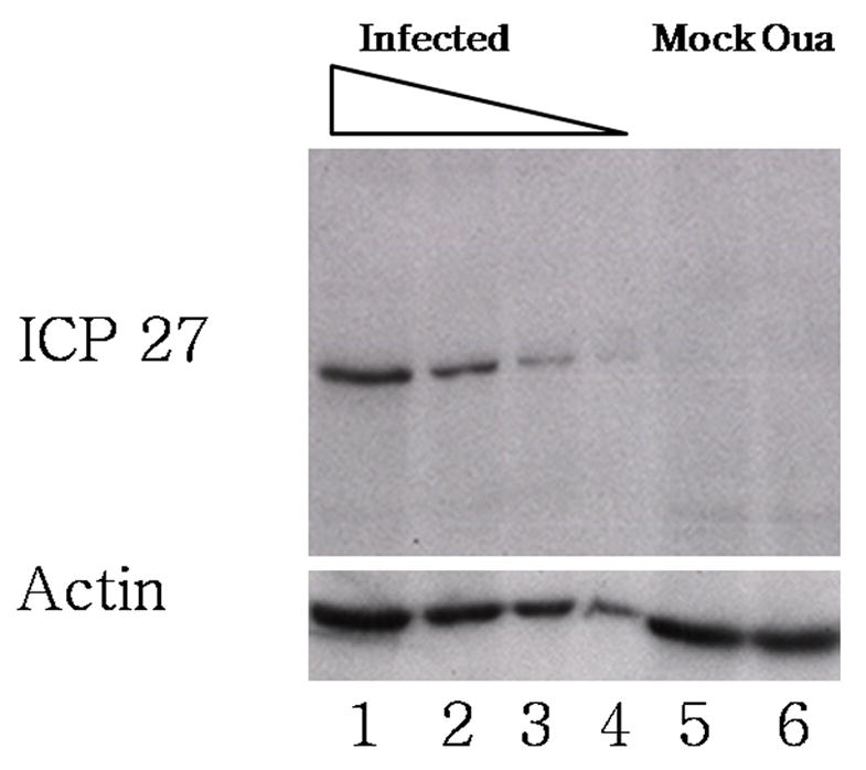
Figure 7.

Effect of ouabain on ICP27 expression. Cells were infected or mock infected in the presence or absence of ouabain. Protein lysates were harvested at 6 hours post infection, and ICP27 was detected by western blot using a monocolonal antibody against ICP27 (top panel). Wells 1–4 contain a series of two-fold dilutions of infected, untreated cell lysate into loading buffer (lane 1 contains undiluted sample, and lane 4 contains a 1:8 dilution). Lane 5 contains undiluted lysate from mock infected cells and lane 6 contains undiluted lysate from infected, ouabain treated cells. The blot was stripped and reprobed using a monoclonal antibody against β-actin (bottom panel).
