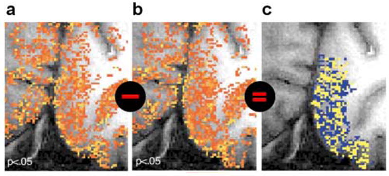
Figure 3. Hi-res fMRI patterns may misrepresent neuronal activity (challenge (1)), but their difference is informative.

Results of Cheng et al. (2001) show that fMRI patterns elicited in V1 by left- and right-eye stimulation (a and b, respectively) are replicable and distinct, but corrupted by vascular artefacts obscuring the ocular-dominance pattern present in the neuronal responses. Because ocular-dominance domains are spatially complementary by definition, they could be visualized by subtracting the fMRI patterns (c). In general, we cannot rely on this assumption and hi-res fMRI patterns will misrepresent the fine structure of neuronal patterns (as seen in a and b). Cheng et al. performed gradient-echo echoplanar imaging at 4T using (0.47mm)2 × 3mm voxels.
