
Fig. 4.
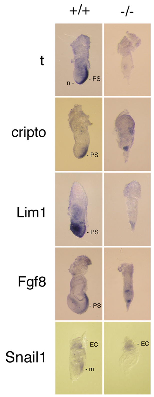
Embryonic gene expression patterning. Embryos were harvested at E7.5 from matings between txnrd1−/+ parents and stained by whole-mount in situ hybridization for the indicated marker genes. After staining and photographing, embryos were digested with proteinase K, nucleic acids were isolated, and genotypes were determined by radioactive PCR (see Fig. 1c). Abbreviations as Fig. 3 except: m, mesoderm; n, node; t, brachyury.