
Fig. 1. FGF9 signaling via mesenchymal FGFR1/2 is necessary and sufficient for distal lung capillary development.
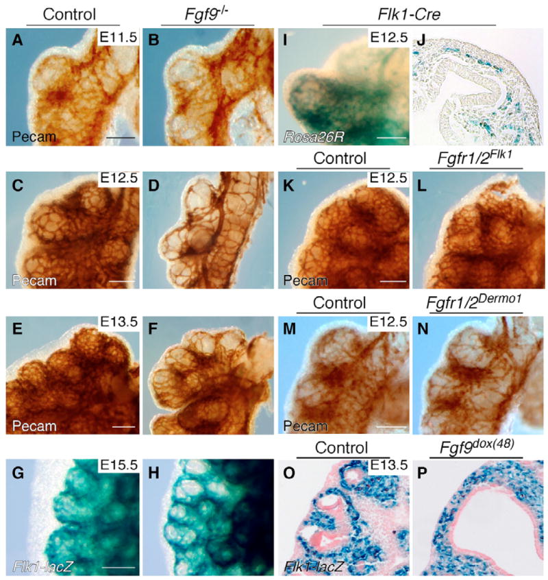
(A–F) Whole-mount immunohistochemistry with anti-PECAM antibody showing large gaps between vessels in the distal capillary plexus of Fgf9−/− lungs at E11.5 (B), E12.5 (D) and E13.5 (F) when compared with control lungs (A,C,E). (G,H) Whole-mount lacZ staining with the endothelial cell marker Flk1-lacZ showing a reduction in capillary density around the distal epithelium in Fgf9−/−; Flk1-lacZ+/− lungs at E15.5 (H) compared with control lungs (G). (I,J) Rosa26R-lacZ stain showing endothelial cell-specific Flk1-Cre activity in a pattern consistent with distal lung endothelial cells in whole-mount (I) and frozen (J) sections. (K,L) Fgfr1 and Fgfr2 double conditional knockout using Flk1-Cre (Fgfr1/2Flk; L) showing no difference in distal lung vascular development compared to an Fgfr1/2f/f control (K). (M,N) Fgfr1 and Fgfr2 double conditional knockout using mesenchymal-specific Dermo1-Cre (Fgfr1/2Dermo1), showing reduced distal lung capillary density (N) compared with an Fgfr1/2f/f control (M). (O,P) Induced Fgf9 expression for 48 hours with doxycycline [Fgf9dox(48)] is sufficient to induce Flk1-lacZ expression throughout lung mesenchyme (P), compared with expression only in the sub-epithelial mesenchyme in control lung (O). Histological sections in O and P were photographed through a 20× objective. Scale bars: 50 μm.