
Fig. 2. Fgf9 is necessary and sufficient for mesenchymal Vegfa expression in the lung.
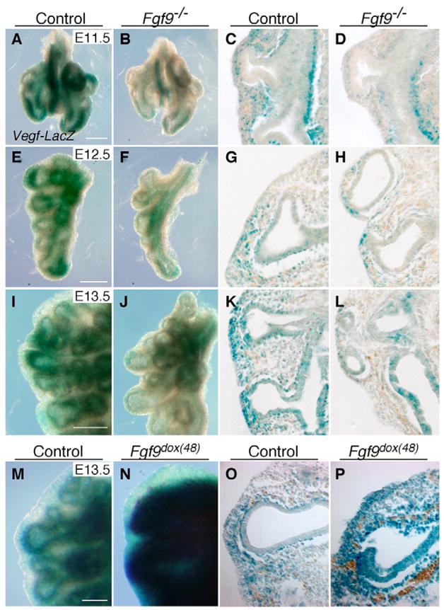
(A–L) Fgf9−/−; Vegfa-lacZ lungs show reduced mesenchymal Vegfa-lacZ staining at E11.5 (B,D), E12.5 (F,H) and E13.5 (J,L) compared with control (A,C,E,G,I,K). At E11.5, no Vegfa is observed in the epithelium (C,D). By E13.5, levels of Vegfa-lacZ in the mesenchyme in Fgf9−/− lungs continue to be reduced compared with controls, but epithelial expression is comparable to controls (K,L). (M–P) Fgf9 overexpression results in increased mesenchymal Vegfa in Fgf9dox(48); Vegfa-lacZ lungs at E13.5 (N,P). Left panels, whole-mount β-galactosidase staining; right panels, cryosections of left panels. Histology: 20× objective. Scale bars: 100 μm.