
Fig. 4. SHH signaling to non-endothelial mesenchyme is necessary for distal lung capillary formation.
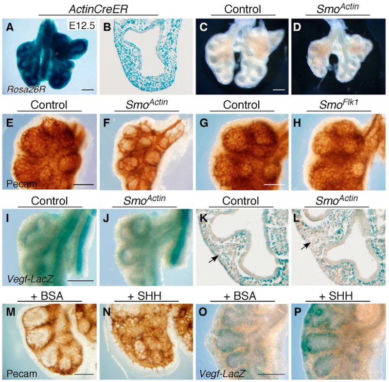
(A,B) The Rosa26R allele was used to detect Actin-CreER activation throughout the lung mesenchyme and epithelium at E12.5 following tamoxifen injection at E9.5; (A) whole-mount and (B) frozen sections. (C,D) Conditional knockout of Smo, using Actin-CreER (SmoActin), results in lung hypoplasia (D) compared with a Smof/f control (C) at E12.5. (E,F) When assessed by anti-PECAM whole-mount immunohistochemistry, SmoActin lungs showed a reduction in distal capillary network density (F) compared with Smof/f controls (E) at E12.5. (G,H) Distal lung capillary density appeared similar in Smof/f; Flk1-Cre (SmoFlk1; H) and Smof/f control (G) mice, indicating that HH signaling to endothelial cells is not required for development. (I–L) SmoActin lungs (J,L) show a decrease in VegfA-lacZ activity in mesenchyme distal to the sub-epithelial layer (arrow) in whole-lung preparations (I,J) and frozen sections (K,L) compared with controls. (M–P) Lung organ cultures incubated with 500 ng/ml of SHH-N protein show an increase in PECAM-positive cells (N) and VegfA-lacZ staining (P) compared with BSA-incubated controls (M,O) after 24 hours. (B,K,L) Lower left lobe; (E–J) upper left lobe. Histology: 20× objective. Scale bars: 100 μm.