
Fig. 5. FGF9 and SHH signaling are both required for capillary formation.
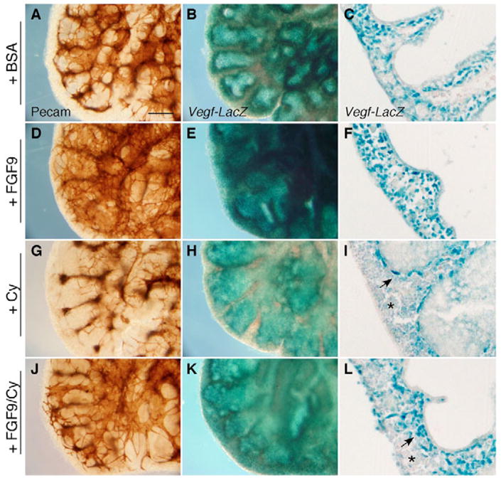
E12.5 lung explants from Vegfa-lacZ mice were incubated for 48 hours with media containing BSA (A–C), FGF9 (D–F), cyclopamine (Cy; G–I) or FGF9 and cyclopamine (J–L). (A,D,G,J) Whole-mount anti-PECAM immunohistochemistry showing increased capillary formation in the presence of 2.5 ng/μl FGF9 (D) and decreased vascular development in the presence of 10 μM cyclopamine (G). FGF9 was able to only partially rescue capillary formation in the presence of cyclopamine (J). (B,E,H,K) Whole-mount preparations and (C,F,I,L) frozen sections stained for lacZ activity. Compared with control explants, Vegfa expression is increased in the presence of FGF9 (E,F) and decreased in the presence of cyclopamine (H,I). FGF9 was able to partially rescue Vegfa expression in the presence of cyclopamine (K,L). Notice that, in the presence of cyclopamine, Vegfa expression is retained only in the inner cell layer of the sub-epithelial mesenchyme (arrow in I), but is significantly reduced in the sub-mesothelial mesenchyme (asterisk in I). FGF9 increases Vegfa expression throughout both mesenchymal layers (F), but in the presence of cyclopamine, Vegfa expression is expanded only in the sub-epithelial layer (arrow in L) and remains low in the sub-mesothelial region (asterisk in L). (A–L) Lower left lobe. Histology: 20× objective. Scale bar: 100 μm.