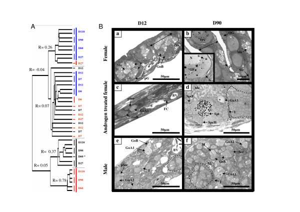
Figure 1.
Classification of gonad samples and histological analysis of some characteristic gonadal stages. (A) Dendrogram of the samples ranked using a hierarchical clustering. Gonad samples are colorized according to the sex i.e., red for females, blue for males and black for androgen-treated females. Correlation coefficients (R) of the last branches of a cluster are given. (B) Histology of the gonads from the female control group (a, b), the androgen-treated group (c, d), and the male control group (e, f) at 12 days (D12) and 90 days (D90) after the beginning of the androgen treatment. BV: blood vessel; FC: fibroblast like cell; Go (A1/A2/B): gonia type (A1/A2/B); I: Interstitial space; M: mitosis; Me: meiosis; N: nucleus; n: nucleolus; OV: ovocyte; P: pachytene stage of meiosis; (p)G: (pre)granulosa cell ; (p)S: (pre)sertoli cell ; Spc(I/II): spermatocyte I/II; Spt: spermatid; Str: ovarian stroma.

