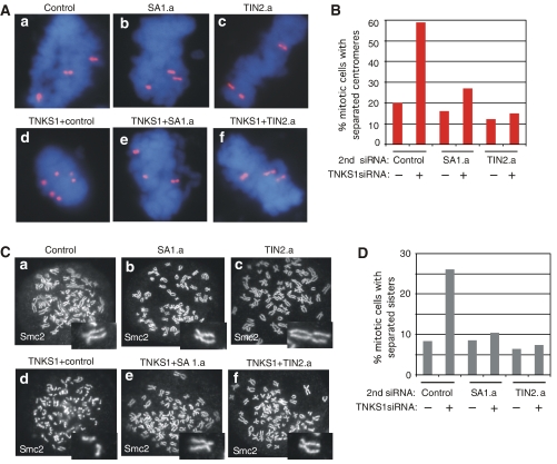
Figure 6.
Depletion of SA1 or TIN2 restores normal centromere cohesion in tankyrase 1-depleted cells. (A) Chromosome-specific FISH analysis of HeLaI.2.11 cells collected by mitotic shake-off at 48 h after treatment with (a) control (GFP), (b) SA1.a, or (c) TIN2.a siRNA without (a–c) or with (d–f) tankyrase 1 siRNA. Cells were fixed directly in methanol-acetic acid without hypotonic swelling and hybridized to a centromere probe 6cen (red). DNA was stained with DAPI (blue). (B) Histogram showing percentage of mitotic cells with separated centromeres; at least 100 mitotic cells were scored for each sample. (C) Chromosome spread analysis of HelaI.2.11 cells collected after 48 h of treatment with (a) control (GFP), (b) SA1.a, or (c) TIN2.a siRNA without (a–c) or with (d–f) tankyrase 1 siRNA. Cells were swollen in hypotonic buffer and fixed in paraformaldehyde. Cells were treated with colcemide for 90 min before harvesting. Chromosome arms were visualized by staining with antibodies to the condensin subunit Smc2. (D) Histogram showing % mitotic cells with separated sisters; at least 200 mitotic cells were scored for each sample.

