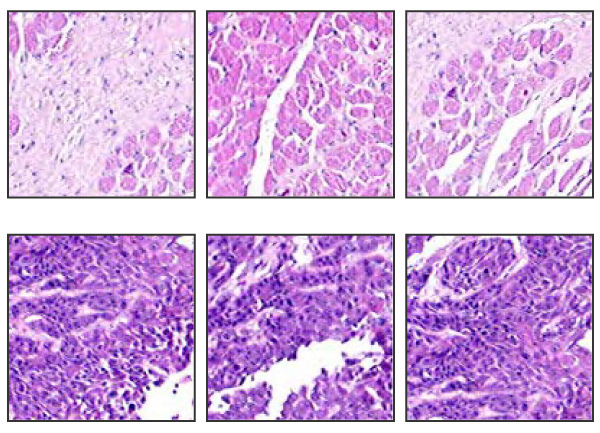
Figure 2.

Three samples from negative (first row) and positive (second row) training subimages. The negative image typically has light red color and low density of cells, while the positive image is characterized by dark color and high density of cells.
