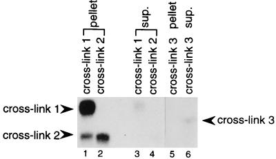
Figure 2.
Immunoprecipitation of the crosslinked species with anti-TMG antibody. Crosslink 1 (lanes 1 and 3), crosslink 2 (lanes 2 and 4), and crosslink 3 (lanes 5 and 6) were excised from gels (Fig. 1B) and incubated with antibody. RNAs recovered from the pellets (lanes 1, 2, 5) and supernatants (lanes 3, 4, 6) were visualized on 5% polyacrylamide gels. The positions of the three crosslinks are indicated. The excised crosslink 1 band was contaminated with a small amount of crosslink 2 (lane 1).

