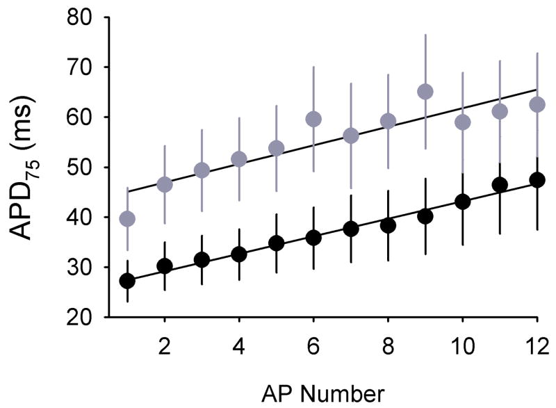
Fig. 4. Action potentials are prolonged in Scn1b null myocytes.
APD75 values were measured in isolated Scn1b wildtype (n=14) and null (n=15) ventricular myocytes in response to stimulation at a cycle length of 150 ms. Solid lines are linear fits to the data. Analysis of the data using two-way ANOVA shows a significant (p < 0.0001) increase in APD75 in Scn1b null myocytes.

