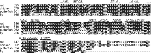
FIG. 1.
Multiple sequence alignment of the SBP2 L7Ae RNA binding domain. SBP2 amino acid sequences from rat, chicken, puffer fish, and fly were aligned. Residues shaded in black are identical, and residues shaded in gray are conservative substitutions. Residues mutated to penta-alanine are indicated above the corresponding positions in SBP2. Sequences were generated using MultAlin, and conserved residues were shaded using Boxshade.

