FIG. 3.
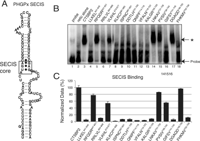
SECIS binding analysis of CTSBP2 penta-alanine mutant proteins. (A) Diagram of the PHGPx SECIS element. The 203-nucleotide PHGPx SECIS element was utilized to assay SECIS binding. The SECIS core motif is boxed to indicate the SBP2 binding site. (B) [35S]Met-labeled wild-type and mutant forms (indicated by the regions corresponding to the mutations) of CTSBP2 were incubated with 20 fmol of the wild-type 32P-labeled PHGPx SECIS element, and the complexes were resolved on a 4% nondenaturing gel. The asterisk indicates the position of a SECIS-specific complex. Unsupplemented reticulocyte lysate (retic alone) was tested as a negative control to account for nonspecific binding. (C) The results presented in panel B were quantitated as the percentage of shifted probe, corrected for nonspecific binding, and normalized relative to the level of shift observed for wild-type CTSBP2. The data shown are the averages ± standard errors of results from at least three independent experiments.

