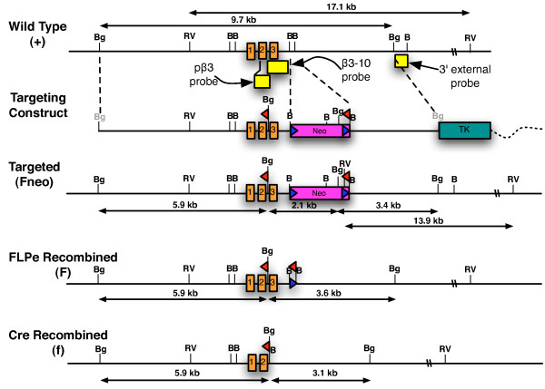
Figure 1.
Gene targeting strategy. Diagram of the wildtype β3 locus illustrating the first three exons (orange boxes) and surrounding genomic DNA (thin, black line). Also shown are the DNA probes (yellow boxes) that were used for Southern blot analysis. The targeting construct included a neomycin (neo) cassette that was flanked by two frt sites (blue triangles), two loxP sites (red triangles), a thymidine kinase (TK) cassette, and plasmid vector backbone (broken, wavy line). During vector construction, the BglII restriction sites shown in grey lettering were destroyed. Also shown is a correctly targeted locus (Fneo), a locus following FLPe mediated deletion of the neo cassette (F), and a locus following Cre mediated deletion of exon 3 (f). Abbreviations: BamHI, B; BglII, Bg; EcoRV, RV.

