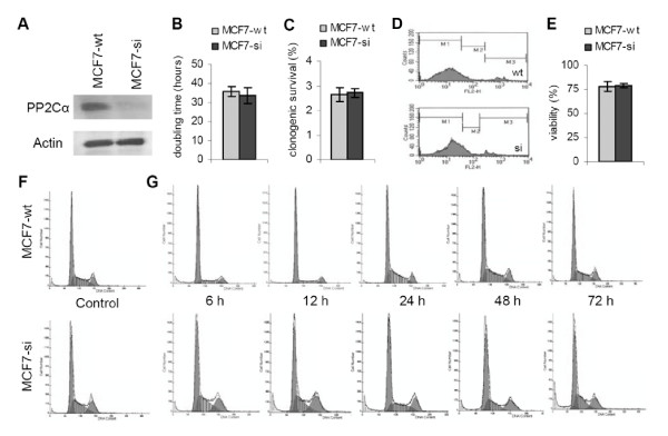
Figure 1.
Characterization of wild type and PP2Cα siRNA-expressing MCF7 cells. A: Western blot analysis of the expression of PP2Cα and Actin in wild type (MCF7-wt) and PP2Cα knockdown (MCF7-si) cells. B: Doubling time of MCF7-wt and MCF7-si cells. Values represent average ± SD (n = 3). C: Clonogenicity of MCF7-wt and MCF7-si cells. Values represent average ± SD (n = 5). D: Representative FACS analyses of the uptake of propidium iodide (PI) by confluently growing MCF7-wt and MCF7-si cells. The uptake of PI was used to determine the membrane integrity of the (unfixed) cells. M1: viable cells. M2: membrane-damaged cells. M3: dead cells. E: Quantification of the viability of MCF7-wt and MCF7-si cells. The amount of viable cells (M1 fraction) was determined by pooling the data obtained in three independent analyses. Values represent average ± SD. F: Representative images of the cell cycle distribution of confluently growing MCF7-wt and MCF7-si cells. G: Representative images of the cell cycle distribution of MCF7-wt and MCF7-si cells at various time points after trypsination and (re-) seeding.

