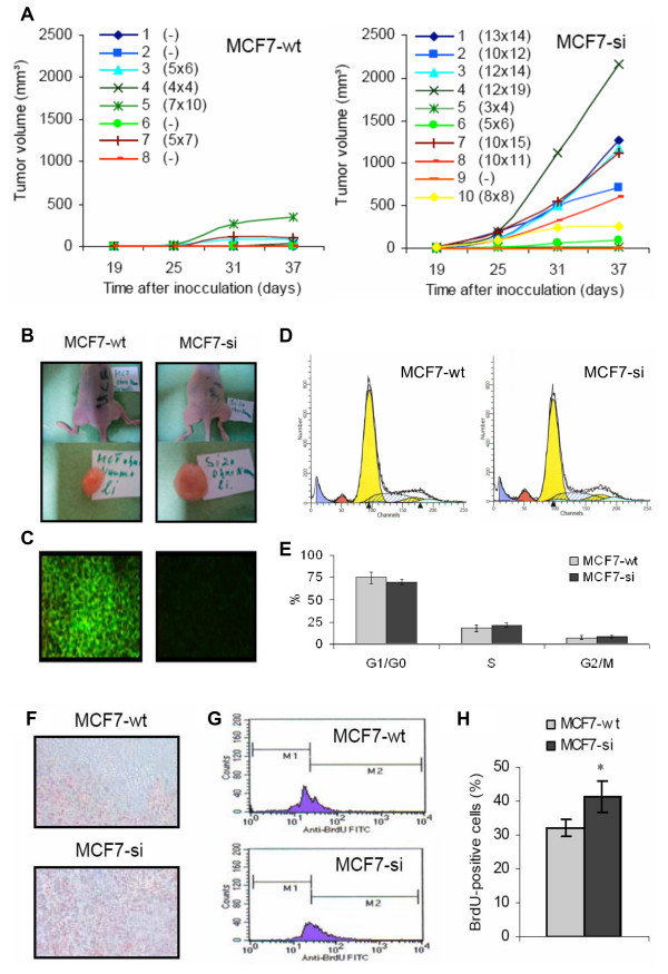
Figure 4.
Evaluation of the tumorigenic potential of wild type and PP2Cα siRNA-expressing MCF7 cells. A: Tumor growth curves obtained for subcutaneously inoculated MCF7-wt and MCF7-si cells upon the combined implementation of estrogen-containing hormone pellets and Matrigel. The values indicate the individual tumor sizes at day 37. B: Exemplary images of MCF7-wt and MCF7-si tumors at day 37 post inoculation. C: Analysis of the expression of PP2Cα in MCF7-wt and MCF7-si tumors. Magnification 200×. D: Representative images of the cell cycle distribution of MCF7-wt and MCF7-si tumors. Left yellow peak: G0/G1 fraction of the aneuploid tumor cells (i.e. the MCF7 cells). Right yellow peak: G2/M fraction of the aneuploid tumor cells. Dashed blue area: S phase fraction of the aneuploid tumor cells. Red peak: G1 fraction of the diploid host cells (e.g. mouse fibroblasts and endothelial cells). Blue peak: Necrotic cells. E: Multicycle algorithm-based quantification of the cell cycle distribution of MCF7-wt and MCF7-si tumors. Values represent average ± SD (n = 3). F: Analysis of the expression of the proliferation marker Ki-67 in MCF7-wt and MCF7-si tumors. Magnification 200×. G: Representative FACS analyses of the incorporation of the proliferation marker BrdU in MCF7-wt and MCF7-si tumors. H: Quantification of the amount of BrdU-positive cells in MCF7-wt and MCF7-si tumors. Values represent average ± SD (n = 3). * Indicates p < 0.05.

