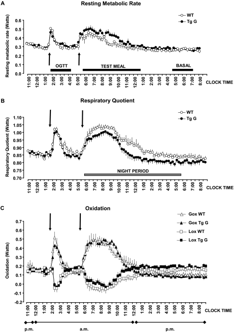
Figure 3. Energy expenditure in mice after impairment of extracellular sugar detection.
Progression of resting metabolism rate, respiratory quotient and calculated oxidation recorded at ten-second intervals in wild-type (WT) and transgenic (Tg) mice. The test meal was given just before the lights were turned off and the response measured until metabolism rate and respiratory quotient returned to pre-meal levels. The changes in resting metabolism rate and respiratory quotient were included in the calculation of the changes in glucose (Gox) and lipid (Lox) oxidation.

