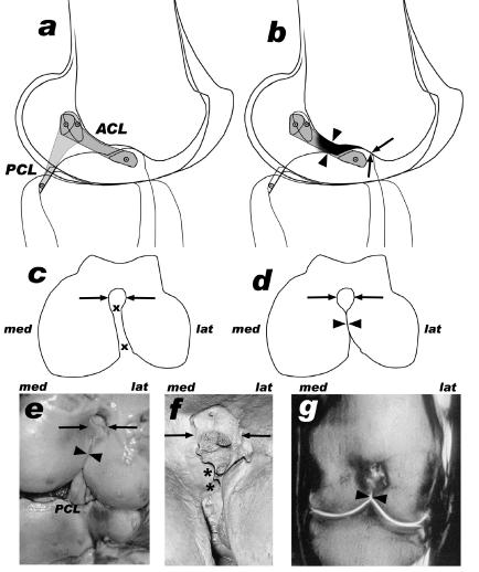
Fig. 6.
Intact (a,c) and arthrotic (b,d–g) knee joints, side view (a,b), distal view of femur (c–f) and dorsal oblique plane (MRI scan, g). In the specimens (both Loxodonta africana) of subfigures (e) and (g) the ACL was missing. Arrows: femoral notch cranial to the Fossa intercondylaris (the corresponding cranial ridge of the tibial intercondylar eminence is in contact with this notch), arrowheads: narrowing of the intercondylar fossa (med = medial, lat = lateral), x–x: passage for the ACL, *: osteophytic ridge.

