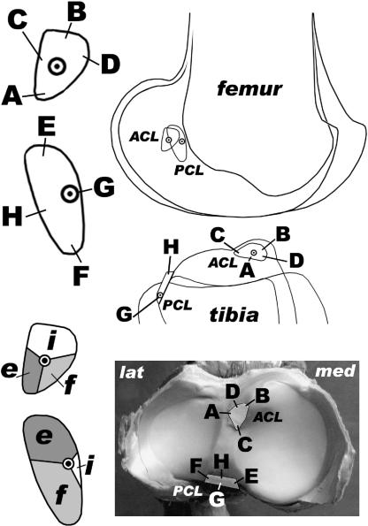
Fig. 9.
Position of the ligament attachments, fibre arrangement and functional subunits. ACL: cranial cruciate ligament, PCL: caudal cruciate ligament, A–D and E–H: four representative fibres of ACL and PCL, respectively. The origin of the isometric fibres is indicated within the footprints. e: fibres taut in maximal extension, f: fibres taut in maximal flexion, i: fibres taut in intermediate positions. Femoral attachments and femur in side view, tibia in side and proximal view (lat = lateral, med = medial).

