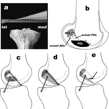
Fig. 11.
Comparison of human and elephant knee. (a) Human helical axis surface [tibia, front view, lat = lateral, med = medial; from Fuss et al. (1997) and Fuss (2001)]. (b) Actual and expected femoral footprints (cf.; Figs 9 and 10), the line x–x indicates PCL fibres which originate from the roof of the intercondylar fossa. (c,d) Human cruciate footprints, four-bar linkage, and shape of the ACL (modified from Fuss, 1989). (e) Arthrotic ridge (arrow) and reduction of the ACL.

