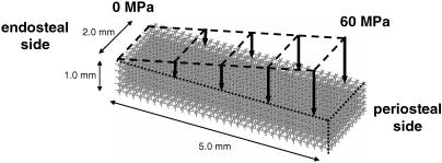
Fig. 1.
The initial finite element model is a 3D rod-like structure. The model was loaded in the longitudinal direction with a distributed load that increased from 0 MPa at the endosteal side to 60 MPa at the periosteal side. The outer part of the model was not loaded to let the model create a natural bone surface.

