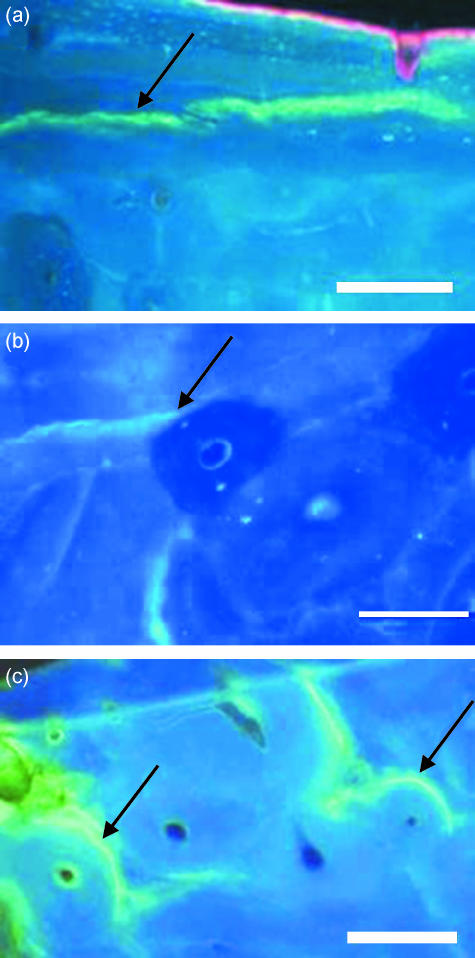
Fig. 1.
Examples of different categories of microcracks, stained with calcein which fluoresces green, shown in transverse sections of bone viewed under UV light (365 nm): (a) microcrack that did not hit any osteon (arrow), (b) microcrack that hit an osteon and stopped outright (arrow), (c) microcracks that hit an osteon and were deflected around the cement line (arrow). Scale bar, 100 µm.

