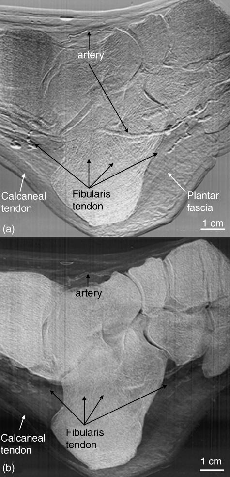
Fig. 3.
Refraction (a) and USAXS (b) images of the same foot as in Fig. 2. Here, a comparison can be made between those structures with edge enhancement from X-ray refraction and those structures having tissues with scatter properties. For instance, in (a), the collagen fibre tissue of the calcaneal and fibularis tendons is readily visible owing to refraction at both fibre bundle edges and the whole tendon edges; these refractile properties differ somewhat from those of surrounding tissue, thus allowing visualization of these structures. Conversely, in (b), the organized parallel structure of the calcaneal and fibularis tendons lack significant scatter properties, and thus X-ray contrast. However, the surrounding fatty and less organized connective tissues display strong USAXS properties. Thus, the distinction between the two tissue types is evident.

