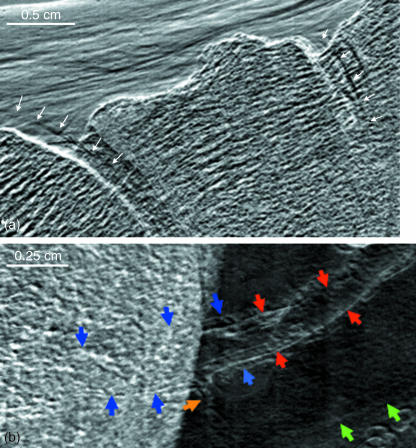
Fig. 4.
(a) A refraction image of talo-navicular and naviculo-cuneiform joints in which the edges of the articular cartilage are visible in regions where they are, and are not, superimposed by bone. (b) Refraction image in which a plantar artery (red arrows) is flanked by venae commitantes (blue arrows). With some limitation, these structures can also be followed through their superimposition by the calcaneal bone. A smaller arterial branch is shown at the yellow arrow. The green arrows point to the inferior border of the tendon of the fibularis longus.

