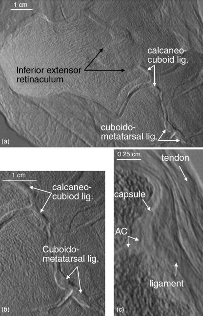
Fig. 5.
(a) A refraction image in which the inferior extensor retinaculum, calcaneo-cuboid and cuboidometatarsal ligaments are readily visible. (b) An enlargement of the calcaneo-cuboid and cuboidometatarsal ligaments. (c) An enlargement of a cuneonavicular joint showing the articular cartilage, joint capsule, ligament and tendon.

