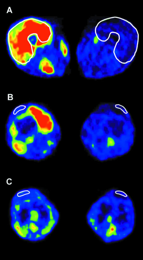
Fig. 2.
Positron emission tomography (PET) images from the regions of quadriceps muscle (A), quadriceps tendon (B) and patellar tendon (C) in the exercising and resting leg of one subject. White lines show the regions of interest. (From Kalliokoski et al. 2005.)

