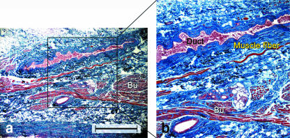
Fig. 5.
(a) Light micrograph of a longitudinal section along the axis of the parotid duct. (b) Higher magnification view of the boxed area in (a) showing distinct muscle fibres originating from the buccinator and running parallel to the duct (Duct). Bu, buccinator. Gomori trichrome. Scale bar = 100 µm.

