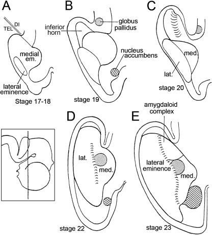
Fig. 8.
The relationship of the ventricular eminences to the parts of the lateral ventricle in dorsal view. (A) In stages 17 and 18 the basal part of the cerebral hemisphere is almost completely occupied by the medial ventricular eminence, whereas the lateral eminence is relatively small. (B) In stage 19 the lateral eminence grows medially and appears to ‘push’ the medial eminence towards the median plane. The inferior horn of the lateral ventricle begins to develop. (C) In stage 20 the inferior horn is better defined. (D) In stage 22 the lateral and medial eminences lie alongside each other and form the floor of the lateral ventricle. The lateral eminence reaches more caudally than the medial and occupies the floor of the inferior horn. The position of the lateral eminence is closer to the definitive topography than is that of the medial eminence. The intereminential sulcus is indicated by short parallel lines. (E) In stage 23 the lateral eminence is larger than the medial. The globus pallidus (stippled) is rostral to the amygdaloid complex. The nucleus accumbens is indicated by oblique hatching. The inset shows the plane of section of D and E.

