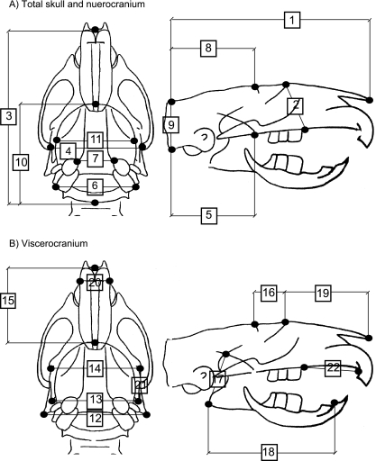
Fig. 1.
Schematic of adult rat skull. (A) Linear measurements of the total skull and neurocranium digitized from the dorsoventral (left) and lateral (right) radiographic views. (B) Measurements of the viscerocranium digitized from the dorsoventral (left) and lateral (right) radiographic views. Measurement numbers and anatomical descriptions of points digitized correspond to those in Table 3.

