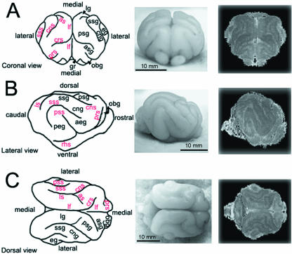
Fig. 1.
Gross anatomical profiles of the ferret brain at 4 weeks of age. Camera lucida renderings (left), photomicrographs (centre), and 3D reconstructions of the 4-week-old ferret brain are displayed in coronal (A), lateral (B) and dorsal (C) views. The prominent, rounded elevations found on the surface of the brain tissue (gyri) with their corresponding acronyms are denoted in black. The furrows along the surface of the brain (sulci) with their corresponding acronyms are denoted in red. The actual sulcal and gyral labels for these abbreviations are listed in Table 2.

