Fig. 1.
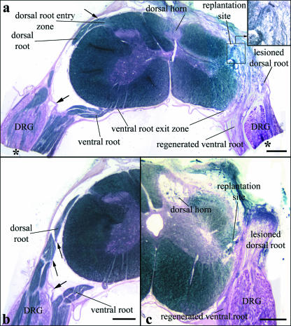
(a) Transverse section of segment C7 with attached dorsal and ventral roots and DRGs 6 months after operation displaying minimal alterations of the dorsal horn on the lesioned side (myelin sheath staining). The distal parts of the DRGs and adjacent ventral roots (asterisks) were embedded in Epon and used to prepare semithin sections for DRG morphological and morphometric analyses (see Materials and methods). The replantation site of the avulsed ventral root and the regenerated ventral root axons are clearly seen (inset). Loose fibrous tissue surrounds the replantation site and extends to the original dorsal root entry zone (DREZ). The dorsal root stump is attached to this fibrous tissue. (b) Morphology of C7 roots and their sheaths on the unlesioned sides. Ventral and dorsal roots are enveloped by thin sheaths, which are in continuity with the spinal cord pia, particularly clearly visible at the dorsal root (small arrows). A thicker sheath, resembling the dura, is interposed between the distal ventral root and the DRG and the dorsal root, separating the two roots completely (large arrows in a and b). (c) Section of C7 in an animal with severe degeneration of the dorsal horn. Scale bars: 500 µm.

