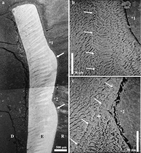
Fig. 5.
Scanning electron micrographs of etched longitudinal sections through lingual enamel of the left M2 of a domestic pig. (a) Two plane-type defects (black arrows) can be seen. Distinct ledges (white arrows) are present cervical to these defects. Note the more obtuse angle between the exposed incremental plane and the ledge in the cuspal (*1) compared with the cervical (*2) defect. D = dentine, E = enamel, R = resin. (b) Higher magnification of the defect marked by *1 in (a). White arrows point to a pathological incremental band. (c) Higher magnification of the defect marked by *2 in (a) and the associated pathological incremental band. Note sharp demarcation of this band (white arrows) against the enamel located internal to it and zone of aprismatic enamel (+) intercalated between the demarcation line and the prismatic enamel located further peripherally.

