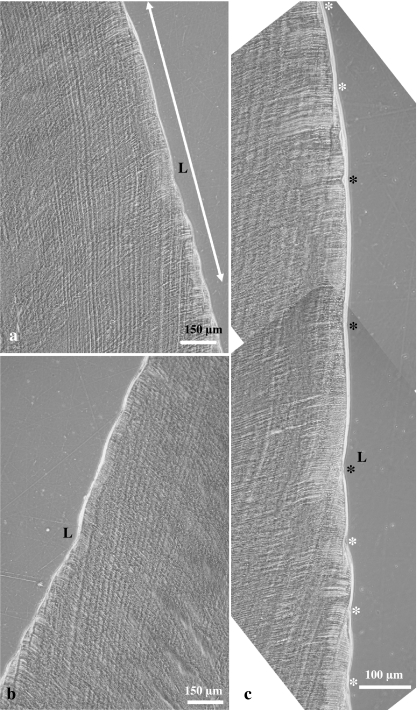
Fig. 6.
Light micrographs (phase contrast microscopy) of a longitudinal ground section through a left M2 of a domestic pig. (a) Linear hypoplastic defect (L) in lingual enamel. The double headed arrow denotes the area magnified in (c). (b) Corresponding linear defect (L) in buccal enamel. (c) Higher magnification of the area marked in (a). Perikyma grooves are marked by asterisks. Note increased distance between three neighbouring perikyma grooves (black asterisks) in the occlusal wall of the defect. White asterisk: normally spaced perikyma grooves.

