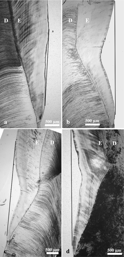
Fig. 7.
Light micrographs (bright-field microscopy) of longitudinal ground sections through lingual enamel of the distal half of M2 from wild boar (a,b) and domestic pigs (c,d). (a) Control molar exhibiting a very slight concavity in the cervical enamel surface. The shortest distance between the deepest point of the concavity of the enamel surface and the tangential line connecting the cuspally and cervically adjacing enamel surfaces is 65 µm. (b–d) Molars exhibiting depression-type defects. Note distinct concavity of both the cervical enamel surface and the course of the DEJ. Maximum depression depth is 491 µm in (b), 388 µm in (c) and 373 µm in (d). D = dentine, E = enamel.

