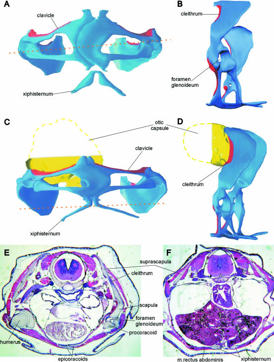
Fig. 2.
Discoglossus pictus. (A,B) Three-dimensional model of pectoral girdle in stage 61. (C,D) Three-dimensional model of pectoral girdle in stage 66. (E) Frontal section at the level indicated by arrows in A. (F) Frontal section at stage 66 at the level indicated by arrows in C. In the models, dermal bones are in red; endochondral elements in blue. A, C in ventral view; B, D, left lateral view.

