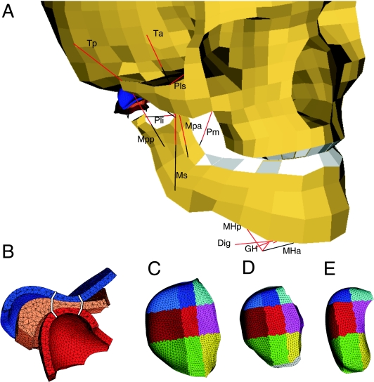
Fig 1.
The model. (A) Anterolateral view. Red lines: muscle contractile element. Black lines: muscle serial elastic element. Ta, anterior temporalis; Tp, posterior temporalis; Ms, superficial masseter; Mpa, anterior deep masseter; Mpp, posterior deep masseter; Pm, medial pterygoid; Pls, superior lateral pterygoid; Pli, inferior lateral pterygoid; Dig, digastric; GH, geniohyoid; MHa, anterior mylohyoid; MHp, posterior mylohyoid. Thin black lines, part of articular capsule. Dig, GH, and Mhp are connected to the hyoid bone (not shown), MHa to the mylohyoid raphe (black line). (B) Sagittal cross-section of the cartilaginous structures of the jaw joint: blue, temporal cartilage layer; orange, articular disc; red, condylar cartilage layer. White lines: separation between anterior, intermediate and posterior regions. (C) Selected regions in the temporal cartilage layer, superior view. Cyan, light blue and blue: medial regions; magenta, light red and red: central regions; yellow, light green and green: lateral regions. Anterior, intermediate and posterior, respectively. (D) Selected regions in the articular disc. (E) Selected regions in the condylar cartilage layer.

