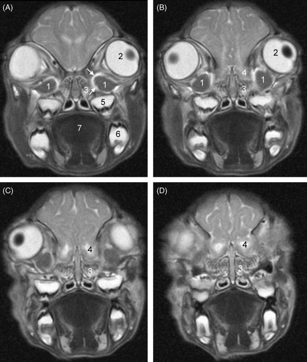
Fig. 1.
T2-weighted oblique coronal MR images through the anterior orbits from posterior (A) to anterior (D). Three- millimetre-thick consecutive slices with 0.3-mm interslice gaps are shown. 1: Harderian glands; 2: left eyeball with (dark) lens; 3: left ethmoidal conchae; 4: left olfactory bulb; 5: tooth, left upper jaw; 6: tooth, left lower jaw; 7: tongue. White arrows: high-intensity rim representing ophthalmic sinus.

