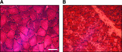
Fig. 2.
The posterior cricoarytenoid laryngeal abductor muscle displays signs of atrophy after RLN transection. Transverse sections of (A) control PCA muscle and (B) PCA muscle 1 month after denervation. Haematoxylin and eosin staining indicates how the mosaic pattern of muscle fibres is lost and the reduction of fibre size after nerve injury. Scale bar = 50 µm.

