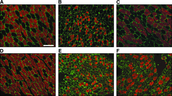
Fig. 4.
Laryngeal muscle fibre morphology after RLN transection. Transverse sections of the posterior cricoarytenoid (top row) and thyroarytenoid (bottom row) muscles immunostained for fast-type MyHC (red) and laminin (green) to highlight individual muscle fibres. A unilateral phrenic–PCA abductor branch anastomosis was performed with repair using PHB nerve conduits (Birchall et al. 2004). Two months after surgery both muscles on the operated side (B and E) showed reduced muscle fibre diameter and expression of fast-type MyHC protein compared with the control side (A and D). After 4 months the PCA muscle fibre diameter appeared similar to control with increased expression of fast-type MyHC protein (C). By contrast, the TA consisted of many small-diameter muscle fibres indicating an atrophic state (F). These results suggest accurate, specific reinnervation of the PCA muscle only. Scale bar = 100 µm.

