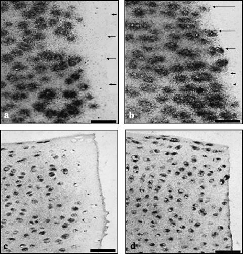
Fig. 6.
Sections showing incorporation of 35S-sulphate (a,b) and 3H-proline (c,d) by wounded explants 2 days after wounding. The trephine wound (a,c), demonstrates a lack of label incorporation adjacent to the wound edge. After wounding with a scalpel (b,d), however, label incorporation is observed adjacent to the wound edge. Scale bar, 50 µm; microautoradiographs counterstained with safranin O (a,b) or haematoxylin and eosin (c,d). Arrows indicate the lesion edge.

