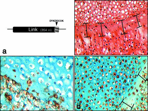
Fig. 7.
Tracking extracellular matrix deposition in fused cartilage using recombinant FLAG-tagged link protein marker. Link protein was cloned with an eight amino acid FLAG-tag linker sequence (a) in order to detect recombinant expression in transfected chondrocytes using anti-FLAG antibodies. Fusion of devitalized cartilage pellet (upper) and overlaid chondrocytes (lower) following 7 days in culture (b). A zone of hypocellularity (indicated) within which are dead cells from the devitalized pellet (black arrow) and filled lacunae (white arrow) are formed at the junction between pellet and overlaid chondrocytes. The same experiment conducted using chondrocytes transfected with recombinant FLAG-tagged link protein shows nascent matrix formation in empty lacunae but limited colonization of the devitalized matrix (c). A combination of live chondrocyte pellet overlaid with transfected chondrocytes expressing recombinant link protein (d) also produces a zone of hypocellularity (indicated).

