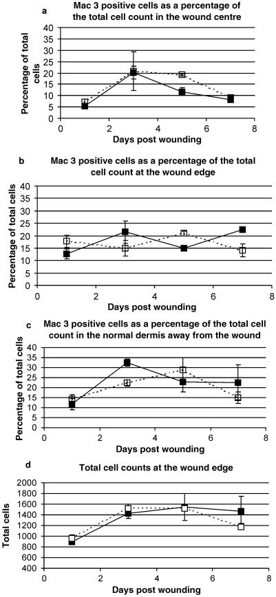
Fig. 8.
Mac-3-positive cell counts expressed as a percentage of the total cell count in both MRL/MpJ (closed boxes) and C57BL/6 (open boxes) wounds. Cells were counted at the wound centre (a), wound edge (b) and in the normal dermis away from the wound (c). There was no significant difference in the percentage of Mac-3-positive cells between the two strains in any region at any time point. Total cell counts at the wound edge (d) demonstrate that no significant difference existed between the two strains of mice, suggesting that percentage cell counts are truly representative. Results shown are mean ± SEM, n = 3.

