
Figure 2.
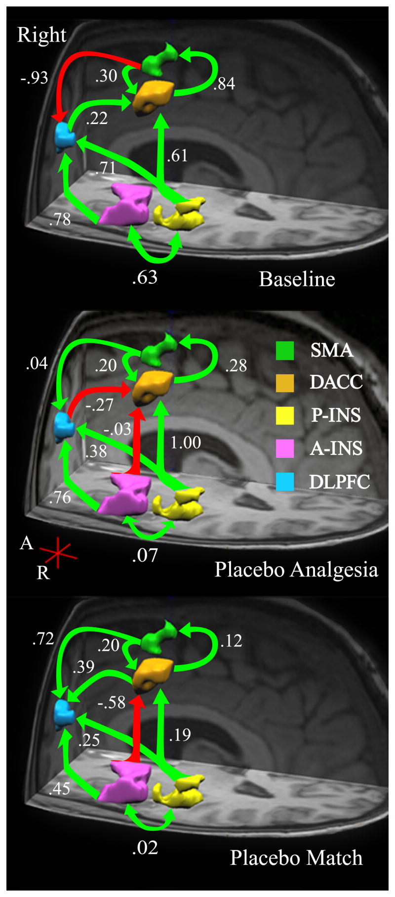
Right hemisphere: Anatomical model of a cognitive – affective pain-related network. Based on a priori hypotheses, brain regions in the right hemisphere (Table 1) were identified a regions of interest (ROIs). The interregional relationships among ROIs are shown here for the left hemisphere across three conditions: Baseline (B1 - top), Placebo (PA – middle), and Placebo Match (PM – bottom). Arrows indicate estimated direction of influence. Green arrows indicate positive path coefficients and red arrows indicate negative coefficients.