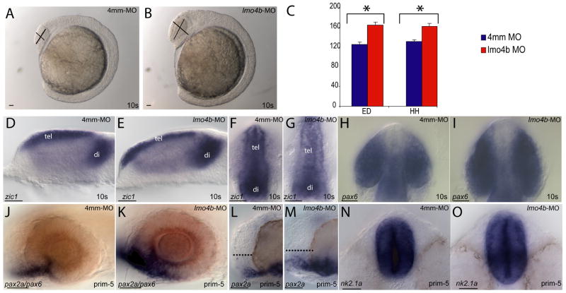
Figure 2. Loss of lmo4b causes forebrain and eye enlargement.
(A, B) Lateral view of 10-somite embryos injected with 4ng of 4mm-MO (A) and 4ng of lmo4b-MO (B). (C) Quantitative representation of eye diameter (ED) and head height (HH) measurements of embryos injected with 4mm-MO (blue) and lmo4b-MO (red). Error bars represent standard error of the mean (SEM). Values in μ with confidence intervals for head height are 131±8 for control injections and 162±12 for morphants, and for eye diameter are 125±10 for control injections and 164±12 for morphants. For HH p= 8.7×10⁻⁵, for ED p= 2.2×10⁻⁵. (D–O) Whole mount views of embryos injected with 4mm-MO (D, F, H, J, L, N) and lmo4b-MO (E, G, I, K, M, O) show size differences in the forebrain and eyes. (D, E, and J–M) are lateral views. (F–I) are dorsal views of the anterior region, and (N, O) are ventral views of the anterior. The dashed lines in (L, M) represent pre-optic area. tel= telencephalon; di=diencephalon. Scale bars are 20μ.

