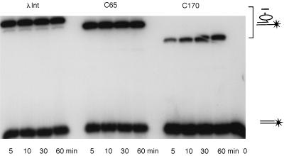
Figure 6.
Covalent complex formation between suicide att site substrate and Int, C65, or C170. One picomol of suicide half att site substrate labeled at the 5′ end of the bottom strand (as illustrated in the right margin) was incubated at 25°C with 20 pmols of intact Int or C65 (Left and Center) or 200 pmols of C170 (Right) in buffer A. At the indicated times, aliquots were quenched with 0.2% SDS, electrophoresed on an 15% SDS/polyacrylamide gel and visualized by autoradiography and quantitated with a PhosphorImager. The cartoon in the right margin indicates the position of the retarded covalent complexes and illustrates the loss of the three-base oligo from the 3′ end of the top strand, thus trapping the covalent complex.

