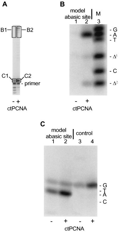
Figure 4.
Determination of which dNMP is recovered opposite the template lesion from oligonucleotide primer extended by pol δ. Incubations were formulated essentially as described in Materials and Methods; as oligonucleotide substrate, they contained the 32P-labeled 38–12-mer shown as oligonucleotide (I) in Fig. 3 (where X = the abasic site) or a similar 38–12-mer where X = dCMP. Because of the relatively short primer, incubations were at 25°C, but for 25 min (A–C) ctPCNA, calf thymus PCNA. (A) After reaction of 32P-labeled 38–12-mer containing the abasic site with proteins as indicated, labeled products were displayed by denaturing PAGE, eluted from gel slices and analyzed further. (B) Fully and near-fully extended products synthesized from 32P-labeled primer were processed according to the strategy outlined in Fig. 3 and subjected to two-phase PAGE exactly according to (47). Lane 1, product eluted from segment B1 of the gel shown in A; lane 2, product eluted from segment B2 of the gel shown in A; lane 3, synthetic oligonucleotide markers subjected to electrophoresis in parallel with reaction products in lanes 1 and 2. (C) Product extended by a single nucleotide was analyzed by standard denaturing PAGE. (Note that this gel system is different from that used in B, thus accounting for the different relative mobilities between B and C, of oligonucleotides with different base compositions.) Lanes 1 and 2, products derived from gel segments C1 and C2, respectively, shown in A; lanes 3 and 4, 32P-labeled 12-mer primer was annealed to a 38-mer template containing a dCMP at position designated X as shown in oligonucleotide (I) of Fig. 3 and extended by pol δ with and without PCNA in the presence of only dGTP. Marker oligonucleotides were subjected to electrophoresis in parallel with the samples loaded in lanes 1–4; their migration positions are indicated to the right of C.

